Image of the month (June 2006)
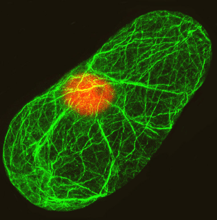
This is a confocal image of an Arabidopsis thaliana cell. This image is from the cover of Plant Journal June 1999 issue, showcasing a representative figure from a paper that appeared in that issue: Kandasamy et al (1999) Plant Journal 18: 681-691.
The text that accompanied the cover succinctly provides both background and explaination for this image. Quoting from the journal:
"Immunofluorescence staining of actin filaments in an Arabidopsis Fi-3 suspension cell with a new plant actin-specific monoclonal antibody, MAbGPa. The ancient actin multi-gene family of Arabidopsis has two major classes, vegetative and reproductive, and five subclasses. To understand the functional significance of the different subclasses of plant actin isovariants, Kandasamy et al. have undertaken the difficult task of producing subclass-specific monoclonal antibodies against the highly conserved actin proteins. In this issue (pp. 681-691), they describe a novel antibody (MAb45a) that distinguishes between the vegetative and reproductive actins at the biochemical and cellular level. The pollen-specific epitope recognized by MAb45a is conserved among diverse angiosperms and advanced gymnosperms like Ephedra, but not lower gymnosperms like Pinus, suggesting that the epitope evolved in an ancestor common to angiosperms and advanced Gymnosperms more than 220 million years ago. The two antibodies characterized in their paper should be useful to plant cell biologists interested in understanding the role of the cytoskeleton in plant development. In the cover image, the actin filaments labeled with MAbGPa (primary) and FITC conjugated sheep anti-mouse antibody (secondary) are shown in green and the nucleus stained with DAPI is shown in orange".
Elegance with which actin filament staining reveals the backbone of a cell is a treat to our eyes. This image also stunningly captures the center of cell universe–nucleus. Another thing I like in this image is that it is one of the few examples that shows how a single Arabidiopsis thalaiana cell looks like. Actin-specific staining, revelation of cell frame work and image acquisition with perfect clarity/focus made this to be our image of the month.
-----------------------------------------------------------------------------
Click here for previously featured image of the month.
|


